
Ultrasound is one of the most widely used diagnostic tools in modern medicine. It allows healthcare providers to see inside the body using sound waves rather than radiation. It makes it both safe and effective. But how do ultrasounds work? At their core, ultrasound machines use high-frequency sound waves to create real-time images of internal organs, tissues, and blood flow. At TrueView Ultrasound, we offer comprehensive ultrasound services that harness this powerful technology to provide clear and accurate diagnostic information for patients across all stages of life.
The Technology Behind Ultrasound
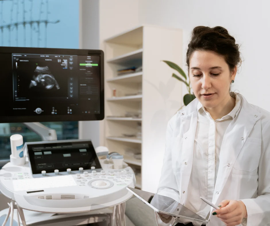
Ultrasound machines operate using a device called a transducer, which emits high-frequency sound waves, typically between 2 to 18 megahertz. These waves travel into the body and bounce off tissues, fluids, and organs. The reflected waves are then received back by the transducer and sent to the ultrasound machine, which processes them into images. The key concept behind how ultrasounds work is echolocation, similar to how bats and dolphins navigate. At TrueView Ultrasound, our advanced equipment captures these sound wave echoes with high sensitivity, producing sharp, real-time images used in diagnostic evaluations.
Different Types of Ultrasound Imaging
Ultrasound technology is incredibly versatile, with several specialized applications. 2D ultrasounds produce flat, two-dimensional images that are typically used for examining internal organs and tracking fetal development. 3D and 4D ultrasounds add depth and motion, providing more detailed visuals, especially in fetal imaging. Doppler ultrasound, another key type, is used to assess blood flow through vessels and detect blockages or clots. Our fetal imaging options at TrueView include 2D to 5D technology. It offers both clinical accuracy and emotionally meaningful experiences for expecting parents.
Why Ultrasound Is Safe and Non-Invasive
Unlike X-rays or CT scans, ultrasound does not use ionizing radiation, making it safer for frequent use, particularly in prenatal care. The sound waves used are well above the range of human hearing and do not pose harm to tissues. According to the Radiological Society of North America, there are no known harmful effects associated with the diagnostic use of ultrasound in human patients. This safety profile is why ultrasounds are commonly used in pregnancy, pediatric care, and routine abdominal or pelvic screenings. At TrueView Ultrasound, we prioritize both precision and patient safety in every exam we perform.
How Ultrasound Images Are Interpreted
Once the sound waves are converted into images, trained technicians and radiologists analyze the results. They evaluate the size, shape, and texture of organs, tissues, or growths, comparing them with established health benchmarks. These insights support the diagnosis of conditions ranging from gallstones and cysts to vascular issues and fetal anomalies. Our staff at TrueView is experienced in interpreting a broad range of scan results and works closely with your referring physician to ensure every detail is reviewed carefully. After your appointment, we deliver accurate reports promptly, contributing to fast and informed medical decisions.
What to Expect During an Ultrasound Exam
Most ultrasound exams are quick and painless. Depending on the type of scan, you may lie on an exam table while a gel is applied to your skin to help conduct the sound waves. The technician then moves the transducer across the area being examined. Some exams, such as transvaginal or transrectal ultrasounds, use a specially designed probe to access internal structures more closely. To make your visit as smooth as possible, our ultrasound preparation guide provides easy-to-follow instructions on what to wear, how to prepare, and what to expect.
Common Applications of Ultrasound in Diagnostics
Ultrasound is used in a wide variety of clinical settings. It is essential in obstetrics, helping monitor fetal development throughout pregnancy. In cardiology, Doppler ultrasounds are used to examine the heart and blood vessels. In abdominal scans, ultrasound can detect gallstones, liver disease, or kidney issues. Thyroid, pelvic, and carotid artery scans are also common uses of ultrasound technology. At TrueView Ultrasound, we provide a full spectrum of diagnostic imaging tailored to each patient’s medical needs, making us a trusted provider of advanced ultrasound services in Roseville.
Conclusion and Next Steps
Ultrasound is a remarkable blend of science and simplicity, harnessing sound waves to visualize the human body in real time. Its safety, versatility, and accuracy make it a cornerstone of modern diagnostics. Understanding how ultrasounds work can help patients feel more informed and confident as they undergo scans for pregnancy, vascular conditions, or internal health evaluations. At TrueView Ultrasound, our commitment to technological excellence and compassionate care ensures that every ultrasound delivers both clinical value and peace of mind.
If you are in need of a diagnostic ultrasound or have been referred by your physician, our team is here to help. TrueView offers state-of-the-art ultrasound imaging in a professional and caring environment. Schedule your appointment today and experience the clarity, precision, and comfort that set our clinic apart.